Day 1 :
Keynote Forum
Shyam S. Mohapatra
University of South Florida, USA
Keynote: Anti-fusion targeted nanomicellar theranostics: Novel antiviral strategies for respiratory syncytial virus infection-induced lung diseases.
Time : TBD

Biography:
Shyam S. Mohapatra is a Distinguished USF Health Professor, Director of the Division of Translational Medicine (Internal Medicine) and Associate Dean of Graduate programs at the College of Pharmacy in the University of South Florida. He also He also directs the USF Center for Research and Education in Nanobioengineering and is also a Research Career Scientist at the James A. Haley VA Hospital in Tampa. He has published over 200 papers and holds over 30 U.S. and foreign patents. He is a Fellow of the AAAAI, NAI, AIMBE and AAAS and a 2014 inductee of the Florida Inventors Hall of Fame.
Abstract:
The respiratory syncytial virus (RSV), is an important pathogen that infects an estimated 64 million people and causes ~200, 000 deaths globally every year. Despite progress in the biology of RSV, there is no effective treatment or vaccine against RSV infection. Currently, only high-risk infants receive antibody-based prophylaxis, which is expensive and moderately effective in reducing hospitalization. Therefore, a broadly applicable, effective and inexpensive approach to prevent or treat RSV-bronchiolitis or -pneumonia remains an urgent unmet need. We have been investigating nanomedical approaches against RSV infection and have reported on a variety of different strategies including genome vaccine, and siRNA based nanoparticles. More recently, we have developed a novel prophylaxis and/or therapy against RSV infection was inspired by the following discoveries. i) A platform of phospholipid micellar nanoparticles (PMN) was developed, which when given intranasally delivers payload predominantly to the lung. ii) A decoy short heptad repeat (HR)2 peptide was identified, which effectively inhibits the RSV-cell fusion. iii) Human mesenchymal cells were found to be highly susceptible to RSV. The latter aided in establishing a novel 3D scaffold for anti-RSV drug screens, which consisted of creating a completely naked mouse lung scaffold(nMLS) by completely decellularization and recellularizing the nMLS with desired human cells such as including hMSCs and epithelial cells and then infecting the cells in scaffold with RSV with or without drugs. iv)
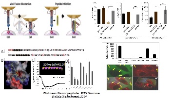
A robust immunocompromised mouse model was created by combining cyclophosphamide treatment with infection by a highly mucogenic strain, RSV-L19F. These developments have led to the hypothesis that a RSV-targeted PMN (RTPMN), combining HR2D anti-fusion peptide, and plasmid encoded siRNAs against RSV-NS1 can provide a safe, effective and inexpensive anti-RSV prophylaxis and/or therapy. The completion of preclinical formulation of anti-RSV PMN-based prophylactics and therapeutics is expected to pave the way to IND-driven studies and clinical trials.
- Nanomedicine and Drug delivery
Location: Novotel Melbourne St Kilda, Melbourne, Australia
Session Introduction
Subhra Mohapatra
University of South Florida,USA
Title: Anticancer Theranostic Approaches for In Vitro and In Vivo Drug Delivery

Biography:
Dr. Subhra Mohapatra is an Associate Professor of Molecular Medicine in the Morsani College of Medicine, University of South Florida. Her laboratory utilizes nanotechnology–integrated cellular and molecular approaches to dissect major signaling pathways in cancers and identify novel drug targets and biomarkers and experimental therapeutics for cancer. Her lab has also synthesized 3D polymeric nano/micro scaffolds for studying tumor-stroma interactions and anticancer drug targets. During last 10 years, she has trained 13 postdocs and 16 graduate students involved in multidisciplinary research, such as biology, immunology, nanoscience and nanotechnology fields. She has authored over 62 scientific papers and holds 9 US patents. She has received many awards for her work including USF-Excellence in Innovation Award. Her research is funded by the National Institute of Health, Veterans Administration and Florida Department of Health.
Abstract:
The last decade has seen significant advances in anti-cancer drug delivery approaches, although many challenges including availability of limited nano- and bio-materials, uptake and release of drugs from the endosomes, targeting of drugs to the desired diseased cells or tissues, and the lack of translatable models to study drug delivery. To address these challenges we have developed and tested a number of novel drug delivery approaches. To this end, we first developed a near infrared (NIR) triggered drug delivery platform based on the chitosan-modified chemically reduced graphene oxide (CRGO) incorporated into a thermosensitive nanogel (CGN). CGN exhibited an NIR-induced thermal effect similar to that of CRGO, reversible thermo-responsive characteristics at 37-42 °C and high doxorubicin hydrochloride (DOX) loading capacity (48 wt%). The DOX loaded nanogel released DOX faster at 42 °C than at 37 °C. Second, since combining chemotherapy with gene therapy has been one of the most promising strategies for the treatment of cancer, we developed a chitosan functionalized magnetic graphene (CMG) nanoparticle platform for simultaneous gene/drug and SPIO delivery to tumor. The results of these studies indicated that CMGs provide a robust and safe theranostic platform, which integrates targeted delivery of both gene medicine and chemotherapeutic drug(s), and enhanced MR imaging of tumors. Further, since gadolinium (Gd) contrast agents that are predominantly used for T1 MR imaging, have high toxicity and potential side effects including nephrogenic systemic fibrosis, we developed an alternative T1 contrast agents, such as Mn for lung imaging. Here we report on the design and synthesis of multifunctional lipid-micellar nanoparticles (LMNs) containing Mn oxide (M-LMNs) for MRI that can also be used for DNA and drug delivery. Finally, we have developed an in vitro model of tumoroid culture platform for testing drug delivery to tumors that closely mimics in vivo tumors. Taken together these advances are expected to lead to better anticancer drug delivery against cancers.
George Altankov
ICREA & Institute for Bioengineering of Catalonia, Spain
Title: Dynamic adhesive environment alters the differentiation potential of young and ageing mesenchymal stem cells

Biography:
George Altankov is ICREA Research Professor in the Institute for Bioengineering of Catalonia. He is a well- recognized scientist in the field of cell-biomaterials interaction and ECM organization. He got his MD in 1974 in Varna Medical Institute, Bulgaria, where also accomplished his PhD (1984). In 1991-1993 he made his postdoc in Southwestern Medical School at Dallas performing studies on the molecular mechanisms of cell adhesion. During his subsequent work in the Bulgarian Academy of Sciences (1985-2005) he grew up to full professor, head of department and deputy Director of the Institute of Biophysics in Sofia. His studies, performed in close collaboration with GKSS Research Center (Germany), were among the first highlighting that tissue compatibility of materials is strongly dependent on the ability of cells to reorganize surface associated matrix proteins, such as fibronectin, vitronectin, fibrinogen and collagen. Other lines of research that boosts his reputation in tissue engineering and nanomedicine are the pioneering studies on the integrin dynamic, cellular interaction with synthetic membranes, nonofibers design for guiding the cellular behaviour. His studies resulting in more than 100 publications in peer reviewed journals and books that are frequently cited.
Abstract:
Engineering dynamic stem cell niche-like environment offers opportunity to obtain better control of the fate of stem cells. We identified, for the first time, that periodic changes in the adhesive environment of human adipose derived mesenchymal stem cells (ADSCs) alters dramatically their asymmetric division but not their ability for symmetric renewal. Hereby, we used smart thermo-responsive polymer (PNIPAM) to create a dynamic adhesive environment for ADSCs by applying periodic temperature cycles to perturb adsorbed adhesive proteins to substratum interaction. Cumulative population doubling time (CPDT) curves showed insignificant decline in the symmetric cell growth studied for up to 13th passages accompanied with small changes in the overall cell morphology and moderately declined fibronectin (FN) matrix deposition probably as a functional consequence of ADSCs ageing. However, a substantial alteration in the differentiation potential of ADSCs from both early and late passages (3rd and 14th, respectively) was found when the cells were switched to osteogenic differentiation conditions. This behavior was evidenced by the significantly altered alkaline phosphatase activity and Ca deposition (Alizarin red) assayed at 3, 14 and 21 day in comparison to the control samples of regular TC polystyrene processed under same temperature settings.

Biography:
Ali Sheikh Bostanabad has completed his PhD from Peoples Friendship University of Russia and postdoctoral studies from Peoples Friendship University of Russia and. He is Research Fellow in Auckland University. He has published more than 15 papers in reputed journals.
Email.id:ALISHKH144@GMAIL.COM
Abstract:
The phenomenon of bioactivity is associated with the formation of acrystallized hydroxyl carbonated apatite (HCA) layer on the bioglass surface, when soaked in a simulated physiological fluid. This layer is similar to the mineral phase of bone. Synthesized bioglasses have been obtained using organic modifiers instead of mineral modifiers, which are the usual precursors for sol–gel synthesis. Hyperthermia treatment is a method of the cancer therapy using the high temperature up to 43ºC which healthy cells survive but tumor cells can’t resist. The materials used to raise the temperature are called as thermoseed and they are ferrimagnetic, ferromagnetic and superparamagnetic particles potentially.
Juan Manuel
Facultad de Ciencias de la UNAM, Mexico
Title: Use and therapeutic application of Nanocarriers (Smart Drugs) to prevention and remediation of Cardiovascular Diseases.

Biography:
J M Vélez is Researcher and Profesor at Escuela Superior de Medicina of Instituto Politécnico Nacional in México City. Nowadays Vélez and his team are working using Nanocarriers with therapeutic application to prevention and remediation of Cardiovascular Diseases in Laboratory Multidisciplinary of Nanomedicine. He has published some papers in reputed journals.
Abstract:
This work focuses on the potential of nanotechnology in nanomedicine, mainly cardiovascular pharmacology discipline, including the highlight rational approaches in design, manufacturing, development, and applications of nanodevices (smart drugs) containing nanoparticles that acts as nanocarriers to controlled and direct for site-specific targeted smart drug delivery into human body using artificial receptors, and unique nanoparticle systems for diagnostics, screening, medical imaging, prevention, and correction of cardiovascular pathologies therapy after administration routes. Our purpose is to offer the most efficient the development pathways for nanomedicine is to merge biomolecular and cellular techniques, tools and method with the nanotechnology knowledge base, as it specifically relates to the development of nanoparticles for enabling and improving targeted delivery of the therapeutic agents; developing novel and more effective diagnostic and screening techniques to extend the limits of molecular diagnostics providing point-of-care diagnosis and more personalized medicine.
Wei Deng
Macquarie University,Australia
Title: Light-triggerable liposomes for enhanced endo/lysosomal escape and gene silencing in PC12 cells

Biography:
Wei Deng received her PhD degree in Chemistry with nanotechnology background at Macquarie University, Australia, in 2012. She was rewarded a highly competitive fellowship (Discovery Early Career Research Award) from the Australian Research Council in 2012. She is now a research fellow in the Centre of Excellence in Nanoscale Biophotonics, Macquarie University. Her research fields were mainly focused on biomedical applications of liposomes and polymer nanoparticles, in particular, light (or X-ray)-controlled drug/gene delivery systems in cancer treatments.
Abstract:
Liposomes are an effective gene/drug delivery system, widely used in biomedical applications including gene therapy and chemotherapy. Here we designed a photo-responsive liposome (lipVP) loaded with a photosensitizer verteporfin (VP). This photosensitizer is clinically approved for photodynamic therapy (PDT). LipVP was employed as a DNA carrier for pituitary adenylyl cyclase-activating polypeptide (PACAP) receptor 1 (PAC1R) gene knockdown in PC12 cells. This has been done by incorporating PAC1R antisense oligonucleotides inside the lipVP cavity. Cells which have taken up the lipVP were exposed to light from a UV light source. As a result of this exposure, reactive oxygen species (ROS) were generated from VP, destabilising the endo/lysosomal membranes and enhancing the liposomal release of antisense DNA into the cytoplasm (Fig.1). Endo/lysosomal escape of DNA was documented at different time points based on quantitative analysis of colocalization between fluorescently labelled DNA and endo/lysosomes. The released antisense oligonucleotides were found to silence PAC1R mRNA. The efficiency of this photo-induced gene silencing was demonstrated by a 74 ± 5% decrease in PAC1R fluorescence intensity. Following the light-induced DNA transfer into cells, cell differentiation with exposure to two kinds of PACAP peptides was observed to determine the cell phenotypic change after PAC1R gene knockdown.

Biography:
Dorota Bociaga is involved in the field of biomedical engineering. In this area she is an author of scientific papers, co-author of two patents and two implementation. Since 2003 coordinates national and international projects mainly concerning use of nanotechnologies in medical applications. Reviewer of scientific papers, expert of medical devices certification and implementation process. In her scientific work she uses the experience gained i.a. at Stanford University, CA (USA, 2012), Join Research Centre, Institute for Health and Consumer Protection, Ispra (Italy, 2005, 2006), Eastman Dental Institute, London (GB, 2004), L’Ecole Catholique d’Arts et Métiers – ECAM, Lyon (France, 2003).
Abstract:
Diamond-like carbon coatings and their modification have been the subject of intense research during recent years. The reason is that the surface coatings can adapt surface properties for special biomedical applications. The upper layer is responsible for an implant’s interaction with surrrounding tissues. Applying the biocompatible DLC coating on the biomaterials surface, the cells reactions can be changed, while the bulk properties of a base material will stay untouched.
The adaption and improvement of the performance and capabilities of DLC coatings can be realized using surface modification technologies. Several deposiotion methods are available. One of them is multi-target magnetron sputtering method which we used. Different concentration of dopants was obtained by changing the magnetron sputtering power during the deposition process. The surface characteristics involved the SEM and XPS analysis as well as the measurement of the surface wettability and surface free energy. The biological assessment of the deposited coatings was based on two complementary cell proliferation and viability assays (live/dead and XTT test) performed using two different cell lines, i.e. EA.hy926 and Saos-2 (ATCC).
The performed research demonstrated that the magnetron sputtering allows to modifiy the metallic implants surface using specific element as a dopant and thus enhance their biological response. Assesment of the surface properties revealed that different elements can improve different properties of the biomaterials. In result the in vitro assessment of the doped DLC coating can suggest its potential best application as implant surface coating.
Hieronim Szymanowski
Lodz University of Technology, Poland
Title: Gradient optical filters for medical applications

Biography:
Hieronim Szymanowski, Professor at the Lodz University of Technology, Institute of Materials Science and Engineering. His main areas of expertise comprise: thin film technology both PE CVD and PVD, optical applications, surface engineering, composite materials. He has authored more than 80 scientific publications.
Abstract:
Low energy light has been continuously gaining importance in medical practice. Its pain-relieving as well as regenerating and microcirculation enhancing activity are well recognized. It has also been shown that polarized light exhibits biostimulating properties.
Light utilising techniques require optical filters with their aim being a removal of unwanted wavelengths from the spectrum emitted by the source. Interference filters are constructed as stack multilayer systems composed of alternated films of high and low refractive index materials. Frequently, low adhesion between these materials causes destruction of the filters, and physical effects on interphase boundaries makes them difficult to manufacture.
This work introduces a novel attitude towards optical filters. A manufacture of filters with a gradient change of refractive index is presented. This gradient results from periodic change of the coating composition, predetermined in the phase of filter design. In the filter realization phase, two materials are deposited. One is silicon dioxide with refractive index of 1.45, while the other comprises silicon nitride with refractive index equal 2.20. Changing their proportions in a continuous and periodic manner results in a gradient periodic change of material refractive index. The technology comprises radio frequency plasma enhanced chemical vapor deposition with a use of tetramethyldisilazane as precursor. A use of nitrogen as a reaction medium leads to silicon nitride coatings, while an application of oxygen results in silicon dioxide films. When the process is carried out in a mixture of nitrogen and oxygen, a material with predetermined value of its index of refraction is deposited.
Anna Sobczyk-Guzenda
Lodz University of Technology,Poland
Title: Gradient optical filters for medical applications.

Biography:
Anna Sobczyk-Guzenda received her PhD degree in the field of Materials Engineering in 2007. She works with thin film deposition using low temperature plasma for numerous applications including health care. Her scientific interests also cover fabrication and modification of composite biomaterials. She is an author of more than 40 publications.
Abstract:
Low energy light has been continuously gaining importance in medical practice. Its pain-relieving as well as regenerating and microcirculation enhancing activity are well recognized. It has also been shown that polarized light exhibits biostimulating properties.
Light utilising techniques require optical filters with their aim being a removal of unwanted wavelengths from the spectrum emitted by the source. Interference filters are constructed as stack multilayer systems composed of alternated films of high and low refractive index materials. Frequently, low adhesion between these materials causes destruction of the filters, and physical effects on interphase boundaries makes them difficult to manufacture.
This work introduces a novel attitude towards optical filters. A manufacture of filters with a gradient change of refractive index is presented. This gradient results from periodic change of the coating composition, predetermined in the phase of filter design. In the filter realization phase, two materials are deposited. One is silicon dioxide with refractive index of 1.45, while the other comprises silicon nitride with refractive index equal 2.20. Changing their proportions in a continuous and periodic manner results in a gradient periodic change of material refractive index. The technology comprises radio frequency plasma enhanced chemical vapor deposition with a use of tetramethyldisilazane as precursor. A use of nitrogen as a reaction medium leads to silicon nitride coatings, while an application of oxygen results in silicon dioxide films. When the process is carried out in a mixture of nitrogen and oxygen, a material with predetermined value of its index of refraction is deposited.
Lukasz Szymanski
Lodz University of Technology,Poland.
Title: Synthesis of magnetic CNTs for cancer treatment.

Biography:
Prof. Dr. Lukasz Szymanski obtaind PhD degree in 2005 and professor in 2016. In 2015 he published book called "Electro-synthesis of carbon nanotubes at atmospheric pressure". He presented research in the field of synthesis of carbon nanotubes using plasma. His research are related to the topic of thermal methods of waste utylisation and synthesis of carbon nanotubes in thermal processes - mainly in reactors using resistive heating and microwave plasma. He is the author or co-author more than 70 publications. He participated in several research projects and now he is a member of the Low-Temperature Plasma Chemistry Commission.
Abstract:
There exist more than one hundred different types of cancer and therefore no particular treatment is offered to people struggling with this disease. There is one promising cancer modality - hyperthermia therapy which is based on exposing body tissues to high temperatures. Carbon nanotubes properties make them more safe in use in medicine and hyperthermy than many other substances. Presently method widely used for Carbon Nanotubes synthesis is the CVD (Chemical Vapor Deposition). It involves the pyrolysis of substances, which contain carbon. The ferromagnetic material located inside the carbon nanotubes may cause heating of them. To do this it is necessary to place the nanotubes in the electromagnetic field. If the carbon nanotube will be connected to a cancer cell can be effectively eliminated. In this paper the CVD furnace with
3 temperature controlled zones for Carbon Nanotubes filled with iron was described. In the first zone the liquid solution of catalyst and gas mixture (Ar + H2) was supplied. The last one was for deposition of carbon nanotubes on silicon wafer. Thanks to characterization of CNTs, it can be stated that the best conditions for synthesis of CNTs are following: infusion speed of catalyst solution should be set between 8.5ml/h and about 9ml/h; gas flow should be fixed at 0.8l/min for hydrogen and 0.5l/min for argon during process of synthesis. Temperature of the first zone should be about 600K and that of the other zones should be 1100K.
Zbigniew Kolacinski
Lodz University of Technology,Poland
Title: Magnetic CNTs for selective ablation of cancer cells.

Biography:
Prof. Dr. Zbigniew Kolacinski has completed PhD, DSc from Lodz University of Technology and received the Professor Title from the President of Poland. He is the leader of the Plasma Technology Group and the author or co-author of more than 250 papers presented at conferences and published in scientific journals. His book “Thermodynamics of short arc plasma” was edited in English and Chinese. He is the member of “High Power Section” and “Plasma Chemistry” of the Polish Academy of Sciences. Prof. Kolacinski is currently working on hyper thermal selective destruction of cancer cells.
Abstract:
The magnetic fluid hyperthermia can be efficient in treating patients with cancer assuming that the magnetic fluid being a colloidal suspension of magnetic nanoparticles is selectively delivered to the tumor site. By exposing the carried particles to an alternating magnetic field a heat energy would be dissipated by the carriers, causing the temperature rise in the tumor’s close vicinity making its ablation. The healthy cells can survive temperatures up to 42°C, but cancer cells undergo apoptosis in therapeutic temperatures of 42 - 45°C.
Carbon Nanotubes (CNTs) are capable of absorbing part of the magnetic field radiation due to van Hove singularities but more effective is to fill them with iron atoms. In this paper we present the results of applying highly Fe doped CNTs as the carriers suspended in buffer fluid creating all together a ferrofluid. However in between the carbon atoms of CNTs, strong van der Waals forces appear. They are the main reason for CNTs’ aggregations in suspensions to occur. Therefore, dispersing CNTs is incredibly challenging. In our case the CNTs were dispersed solely in gelatine or in gelatine with SDS (sodium dodecyl sulphate). The fluid was subjected to hyperthermia heating, as well, to simulate the reaction of magnetic CNTs in an alternating magnetic field of radio frequency. The temperature growth characteristic curves will be presented and discussed.
The tests performed on CNTs ferronanofluids have shown that it is possible to obtain required heat dissipation in cancer cells.
Olga Pechanova
Slovak Academy of Sciences,Slovak Republic.
Title: Protective effect of nanoparticle-loaded aliskiren on aortic structure during hypertension.

Biography:
Will be updated soon.
Abstract:
Aliskiren is the most recent antihypertensive agent that acts by inhibition of renin, the first step in renin−angiotensin−aldosterone-system. Aliskiren has been shown to exert renoprotective, cardioprotective, and anti-atherosclerotic effects independent of its blood pressure (BP) lowering activity. Clinical use of aliskiren is limited, however, by short lifetime of this drug. Therefore, the aim of our study was to determine the effect of nanoparticle-loaded aliskiren, with gradually realized drug, on BP and structural alterations of the heart and aorta developed due to hypertension. 12-week-old male SHRs were divided to the untreated group, group treated with powdered aliskiren (25mg/kg per day), group treated with nanoparticle-loaded aliskiren (25mg/kg per day), and group treated with nanoparticles only for 3 weeks by gavage. BP was measured by tail-cuff plethysmography. Collagen and elastin contents were determined by picro-sirius red staining in both heart and aorta. Wall thickness (WT), inner diameter (ID) and cross sectional area (CSA) were determined in the aorta. At the end of experiment, BP was lower in both powdered aliskiren and nanoparticle-loaded aliskiren groups with more pronounced effect in the second one. Moreover, nanoparticle-loaded aliskiren was able to decrease collagen content (by 11%) and CSA (by 25%) in comparison to the powdered aliskiren group, while it had no significant effect on the similar parameters in the heart. There were no significant changes in elastin content, WT and ID among aliskiren groups and control group. Polymeric nanoparticles, however, increased collagen and elastin contents and WT of the aorta. In conclusion, nanoparticle-loaded aliskiren seems to be promising drug in large vessels protection, more suitable polymeric nanoparticles, however, are needed for better tissue protection.
Supported by grants, APVV-0742-10, APVV-14-0932 and VEGA: 2/0195/15, 2/0144/14.
Yaping Li
Chinese Academy of Sciences,China.
Title: Intelligent Nanoparticles for Combination Photoimmunotherapy of Cancer.

Biography:
Prof. Yaping Li received Ph.D. degrees in Fudan University in 2001. He devoted himself in drug targeted delivery based on nanotechnology, mainly involved in reversing MDR and improving pharmacological efficacy of antitumor agents, designing and constructing new non-viral vector for gene delivery. He has published over 150 scientific papers in Nat Medicine, Adv Mater, ACS Nano, Adv Funct Mater, Small, Biomaterials, Nanomedicine NBM, J Control Release, etc. He won National Science Fund for Distinguished Young Scholars (2009), Shanghai Outstanding Academic Leaders (2011), Zhu Li Yuehua Excellent Teacher Award of CAS (2010), the Hundred Talents Program Talent of CAS (2010) and Shanghai Leading Talent (2010). He is the Chief Scientist of National Basic Research Program of China in Nanoscience and Nanotechnology (2009), Vice Chairman of Pharmaceutics Society of Shanghai Pharmacy Association, Member of Council, Shanghai Pharmaceutical Association, Member of Branch of China in the International Controlled Release Association (CRS), and Member of the Chinese Pharmaceutical Associstion.
Abstract:
Photoimmunotherapy (PIT) has emerged as a promising clinical modality for cancer therapy due to its ability to initiate an antitumor immune response. However, PIT is severely impaired by tumor cell immunosuppression of host T-cell antitumor activity through the programmed cell death 1 ligand (PD-L1) and programmed cell death receptor 1 (PD-1) (PD-L1/PD-1) immune checkpoint pathway. In this study we demonstrate that PIT can be augmented by PD-L1 knockdown (KD) in tumor cells. We rationally designed a versatile micelleplex by integrating an acid-activatable cationic micelle, photosensitizer (PS), and small interfering RNA (siRNA). The micelleplex was inert at physiological pH conditions and activated only upon internalization in the acidic endocytic vesicles of tumor cells for fluorescence imaging and PIT. The combination of PIT and PD-L1 KD showed significantly enhanced efficacy to inhibit tumor growth and distant metastasis in a B16-F10 melanoma xenograft tumor model. These results suggest that acid-activatable micelleplexes utilizing PDT-induced cancer immunotherapy are more effective when combined with siRNA-mediated PD-L1 blockade.
Fei Tang
Tsinghua University, Beijing,China
Title: Novel mussel-inspired Ti-6Al-4V surfaces with biocompatibility, blood ultra-drag reduction and superior durability

Biography:
Fei Tang received Dr. degree from Tsinghua University in 2003. He is an Associate Professor in Tsinghua University. He is currently the Deputy Secretary-General of the China Society for Micro-nanotechnology and the Director of the Office. He is a guest editor of the international journal AIP Advances, and mainly engaged in material testing and analysis technology and equipment, micro-system design and technology, precision measurement and control technology, mechanical and electrical integration technology research.
Abstract:
In order to develop new Ti-based biomaterials with biocompatibility, blood ultra-drag reduction and superior durability, a novel fabrication combining simple electrochemical and chemical processes was proposed. After being modified by C14H19F13O3Si (FAS), a biocompatible TiO2-SiO2-polydopamine composite surface on Ti-6Al-4V substrate was obtained. The biocompatibility was evaluated using a series of in vitro test, revealing that compared with Ti-6Al-4V alloys, the surfaces exhibited a number of bio-advantages such as anti-platelet aggregation, anti-bovine serum albumin protein adsorption, a lower hemolysis rate (~0.7 %) and non-cytotoxicity (the cell viability >88%). The test of human microvascular endothelial cells (HMEC) cultured on the specimens for 48h showed better cell proliferation of the surface. Moreover, we explored the blood dynamic characteristics of titanium alloy substrate biomaterial for the first time, with a focus on the effects of dopamine-reactant concentration on blood flow resistance. The results showed that, compared to titanium alloy material, the TiO2-SiO2 surface modified by 4 mg mL-1 dopamine solution displayed the optimal blood drag reduction characteristics, reaching a 76 % drag reduction. After a 2 m (800 meshes, 3500 Pa) sandpaper abrasion test, the surface still maintained a superior repellency of blood (contact angles> 150°, sliding angles < 10°). This practical method may expand the applications of biomedical implantation materials.
Jun Chen
Fudan University,China
Title: Microwave synthesis of Near Infrared Type II water-soluble Lead Sulfide Quantum Dots For in vivo Bioimaging

Biography:
He is currently the assistant Professor of Huashan Hospital, Fudan University, China. He obtained the doctor degree of biomedical engineering from Shanghai Jiao Tong University in 2013, he has had studied in the Central of Bioscience in University of Nottingham as an exchange student from April to Dec, 2013. He moved to Huashan Hospital, Fudan University for two years as a postdoctor, and then leads a multidiscipline research group on nanomateirals and biomedicine. Until now, He has had published more than 20 articles in high impact factor international journals such as Chem. Mater., Biomaterials, Nanoscale, J. Mater. Chem. B, and Chem. Commun., etc, and also has 7 Chinese patents of invention. His research interests including tissue engineering, nanotechology, molecular imaging and multifunctional materials for biomedical application.
Abstract:
Ribonuclease-A (RNase-A) encapsulated PbS quantum dots (RNase-A@PbS Qdots) which emit in the second near-infrared biological window (NIR-II, ca. 1000−1400 nm) are rapidly synthesized under microwave heating. Photoluminescence (PL) spectra of the Qdots can be tuned across the entire NIR-II range by simply controlling synthesis temperature. The size and morphology of the Qdots are examined by transmission electron microscopy (TEM), atomic force microscopy (AFM), and dynamic light scattering (DLS). Quantum yield(QY) measurement confirms that the prepared Qdots are one of the brightest water-soluble NIR-II emitters for in vivo imaging. Their high QY(∼17.3%) and peak emission at ∼1300 nm ensure deep optical penetration to muscle tissues (up to 1.5 cm) and excellent imaging contrast at an extremely low threshold dose of ∼5.2 pmol (∼1 μg) per mouse. Importantly, this protein coated Qdot displays no signs of toxicity toward model neuron, normal, and cancer cells in vitro. In addition, the animal’s metabolism results in thorough elimination of intravenously injected Qdots from the body within several days via the reticuloendothelial system (RES), which minimizes potential long-term toxicity in vivo from possible release of lead content. With a combination of attractive properties of high brightness, robust photostability, and excellent biocompatibility, this new NIR-II emitting Qdot is highly promising in accurate disease screening and diagnostic applications.
Zifu Li
Huazhong University of Science and Technolog,China
Title: Hydroxyethyl starch (HES) based smart nanomedicine.

Biography:
Zifu Li obtained his PhD in 2012 from the Chinese University of Hong Kong. Afterwards, he worked as a postdoctoral in University of Alberta from 2013 to 2015 and as a research scientist in Georgia Tech from 2015 to 2016. He is currently a professor in National Engineering Research Center for Nanomedicine, College of Life Science and Technology at Huazhong University of Science and Technology, Wuhan, China. He has focused on the study of Hydroxyethyl starch (HES) based smart nanomedicine for cancer immunotherapy and combinational therapy.
Abstract:
Hydroxyethyl starch (HES) is a semi-synthetic polysaccharide and has wide clinical use as plasma volume expander. HES is synthesized from waxy maize, which contains more than 95% of amylopectin. HES is highly water soluble and keeps the branched structure of amylopectin. HES can be categorized into various classes based on its molecular weight, mole substitution of hydroxyethyl, and substitution pattern (C2/C6 ratio). These parameters affect the α-amylase-mediated degradation of HES in blood, thus determine the pharmacokinetics of HES, enabling very convenient ways to tailor the in vivo fates of HES by simply adjusting these parameters. The good manufacturing practice, high water solubility, tailorability, biocompatibility, biodegradability, well defined in vivo safeties, and wide clinical applications make HES a promising drug carrier which warrants clinical translation explorations.
In this talk, we will present our recent progress of HES based smart nanomedicine. We prepared a novel redox-sensitive hydroxyethyl starch-doxorubicin conjugate, HES-SS-DOX, with diameter of 19.9 ± 0.4 nm, to alleviate the side effects and improve the antitumor efficacy of DOX. The redox-sensitive HES-SS-DOX has been proved an effective and safe prodrug of DOX for cancer chemotherapy and could be potentially translated for clinical trials. In fact, our HES-based tumor microenvironment-sensitive prodrug strategy is ready applicable to a wide range of drugs, like PTX, Docetaxel, and 5-Fu, for various cancers.
Hawraz Mohammed Amin
University of Salahaddin-Erbil/Iraq.
Title: Cytotoxic effects of bioactive compounds isolated of Iris persica L. on human cancer cell lines

Biography:
Abstract:
Genus Iris (Iredaceae) comprises over 300 species; 12 of them are present in Iraq. Iris persica has been used in Kurdish traditional medicine for the treatment of wound inflammation and tumor. However, Chemical and biological aspects of I. persica have not yet been investigated. The present study reports the first investigation on the isolation and characterisation of bioactive compounds from flowers, bulbs and rhizomes of I. persica that has been collected from Kurdistan Region-Iraq and cytotoxicity effect of the isolated compounds against six human cancer cell lines were evaluated.
Dry flowers, bulbs and rhizomes of I. persica were exhaustively extracted by maceration at room temperature, solvents of increasing polarity: hexane, methanol, methanol/water 70:30. Chlorophylls were removed from the methanolic extracts of flowers by filtration on a C-18 reversed phase column. Subsequently, the methanolic extracts of the flowers, bulbs and rhizomes were separately fractionated by repetitive preparative MPLC, on C-18 reversed phase, affording four compounds as the major products: tectorigenin (1), embinin (2), isovitexin (3) and trans-resveratrol-3-O-β-D-glucopyronoside (4). The structures of the compounds were identified on the basis of spectroscopic analyses and comparison with literature data.